A Brief Essay on Anatomical Drawing
A Brief Essay on Anatomical Drawing
by Hal Sharp
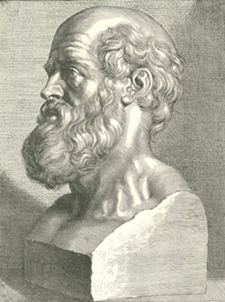
Traditional portrait-bust of Hippocrates Lint, p. 19.
Comparative Anatomy in Classical Greece
“Anatomy is the foundation of medicine,” the classical Greek physician Hippocrates declared, “and should be based on the form of the human body.”(Persaud, p. 33) Certainly no faculty member of a modern medical school would contradict the ancient master on this point, yet for many centuries – indeed for millennia – anatomical study had a distinctly uneasy relationship with medical education and practice. Aristotle, the famous natural philosopher two generations younger than Hippocrates, also considered anatomical study critical to medical knowledge and practice, but he developed theories of human anatomy based only on exterior physical examination and the dissection of animals. In this important work lay the foundations for comparative anatomy, still it proved inadequate to explain the particularities of the human body. Among Aristotle’s many students was the young Alexander, son of Philip of Macedon, and when Alexander earned the epithet “the Great” by conquering a vast territory in the third century B.C. he made Greek intellectual achievements a dominant force in the civilized world. The Greek project found perhaps its greatest expression in the Library of Alexandria, in the city Alexander established on the Nile delta. Scholars and students of many disciplines congregated there and examined both Aristotle’s comparative anatomical studies and the works of Hippocrates.
The Hippocratic Corpus catalogued in Alexandria’s renowned library was a collection of works by Hippocrates himself and successor physicians practicing at his school on the eastern Aegean island of Kos.
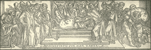
Galen dissecting a pig
In the fifteenth and sixteenth centuries a revival of interest in Galen’s work resulted in Latin translations illustrated by imaginary scenes. Shown here is the dissection of a swine with more animals being bundled in on the right. Lint, p. 22.
Hellenistic Greece and Human Dissection
The availability of the Corpus in the stimulating intellectual environment of Hellenistic Alexandria likely inspired medical scholars finally to take up the knife and undertake systematic human anatomical studies for the first time in classical history. One of these physician-scholars was Herophilus, who produced the treatise On Anatomy, synthesizing the results of many hundred human dissections. It seems probable that such thorough examinations must have been accompanied by drawings, though none have survived, and none of the classical works existing in transcription mention Hellenistic anatomical drawings.
Rome and Anatomical Reversal
With the conquest of Alexandria by Rome, a significant detriment to the advance of medicine occurred in the reversal of the cultural climate favorable to human dissections. Although the great Roman physician Celsus, of the first century AD, and Galen, of the second, both refer to Alexandrian scholarship, the resurgent taboo on human dissection coupled with the loss of the text On Anatomy allowed descriptions of human anatomy presented as fact to slip back into the untested – and largely incorrect – domain of theory.
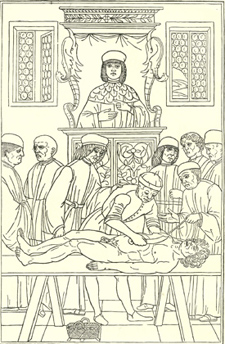
This anatomical demonstration shows the professor in full robes overseeing a dissection and an assistant poised to make his incision. Lint, p. 34.
The Islamic Golden Age and Anatomy
The West owes a great debt to the Arab civilization which conquered the Byzantine successors of Ancient Rome. While much of the western world lost touch with the classical past, Islamic scholars collected and translated the works of Hippocrates and Galen – among many others – and absorbed and synthesized the ancient theories with new discoveries of their own. Despite advances in medical scholarship, Quranic injunctions against human dissection and human representation in drawing placed severe limitations on the critical and accurate analysis of human anatomy. Later in the Medieval period, the resurgence of Europe led to the establishment of universities and the retranslation of classical works into Greek and Latin. In the fourteenth century, some rare human dissections were carried out at European medical schools, ordinarily with the anatomist seated on a dais at the head of a large room, reading from a manuscript of Galen, while a demonstrator opened the body, surrounded by students and other scholars.
Modern Anatomy in the Renaissance
It is with the Renaissance, however, that the era of modern anatomical studies begins. A number of developments united at this time to form a genuinely new cultural system that produced revolutions in many disciplines. The invention of the printing press in the mid-fifteenth century supplanted the laborious process of hand copying and illumination, and made possible the comparatively rapid and widespread dissemination of knowledge in the form of identical books with wood-cut prints. The development of perspective in art led to much greater accuracy in the depiction of individuals and scenes, and prompted artists to make careful studies of human anatomy – including dissections – to educate themselves in the precise structure and musculature of the body. Finally, intellectual attitudes began to break away both from the rigid structures imposed by religious doctrine and by uncritical acceptance of ancient authorities – not everywhere, of course – but the empirical testing of theories by observation and experimentation lay the foundations for the scientific advances and documentation of knowledge that characterized the eighteenth-century enlightenment and our own modern scientific method.
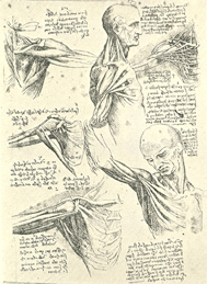
Studies of the shoulder and neck by da Vinci. Lind, p. 39.
In terms of Renaissance anatomy, the two names most closely associated with the progression toward critical analysis and accuracy are Leonardo da Vinci and Andreas Vesalius. Da Vinci approached anatomy from the point of view of the artist, but his meticulous and inquisitive mind quickly propelled him into an anatomical project for its own sake, unfortunately never published. His more than 750 anatomical drawings – most made from observations of dissections he performed – carried the genre of anatomical illustration to extraordinary refinement.
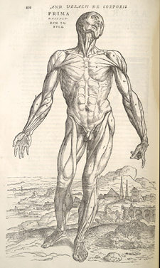
Vesalius’s Fabrica is a masterpiece of accuracy and a work of extraordinary beauty as well. Vesalius, p. 210.
Vesalius, born in 1514, five years before the death of da Vinci, studied medicine in Paris, Louvain (Belgium) and Padua (Italy), where the faculty named the 23-year-old a Professor of Surgery and anatomy at the completion of his doctorate. Vesalius made a name for himself among his students by descending from the chair to conduct dissections without an intermediary, and earned renown among the faculty and profession by successfully challenging the authority of Galen. He put the ancient master to the critical test and revealed errors in his anatomical theory. A series of six anatomical drawings produced by Vesalius proved so popular with his students that he published them in 1538, and here likely germinated the idea for his masterpiece, De Humani Corporis Fabrica, first published in 1543. This volume paired detailed descriptive text with carefully worked anatomical drawings, both of details and entire bodies, all rendered with extreme accuracy, not the least emphasized by the external supports given the sequence of standing figures who successively lose the capacity to “stand” on their own as the layers of musculature are removed. In the perspective distance behind most of these figures is a continuous cityscape of Padua, a further subtle statement of accuracy or truthfulness in the illustrations, as well as an indication of the important scholarly environment within which Vesalius worked. The frontispiece of the Fabrica likewise shows Vesalius himself at the dissecting table, reinforcing his position as a scholar not hidebound by conventional thinking.
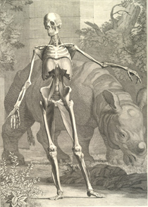
Albinus and artist-engraver Jan Wandelaer took over 8 years to complete this remarkable anatomy atlas with its aesthetic style, scientific accuracy, and fanciful backgrounds. Albinus, table 4.
From this point forward, many, many new anatomical titles appeared, some overtly plagiarized from Vesalius, others new studies, but human dissections and the goal of accurate anatomical illustration never again fell away from the medical curriculum.
Progress in Printing Impacts Anatomy
By the eighteenth century, advances in printing technology enabled the production of larger, folio volumes with copper plate engravings of even greater clarity of detail over the earlier wood cut blocks. The most exceptional author of this era is Bernhard Siegfried Albinus, whose copper plate engravings derived not from freehand drawings wholly dependent on the eye of the artist but from an elaborate surveying mechanism used by architects that regulated artistic talent to make accurate, verifiable measurements and produce precisely proportioned details.
The nineteenth century brought further technological advances in printing, particularly chromolithography, a method for accurate color printing, and changes in paper production methods that lowered costs. These permitted an even greater diffusion of images and texts, making single books and prints available not simply at the level of the school or the professor, but also that of the classroom, student, and practitioner. One type of publication in use at the end of the century was the anatomical chart which brings us to the inspiration for this exhibit: to highlight the anatomical charts in the Claude Moore Health Sciences Library at the University of Virginia.