“Numbers too large to be accommodated at lecture with such a regard to health or comfort as prudence demands” require separate lecture rooms. June 28, 1850. [1]
Creating Room for Classes, the Boiling House and the Dissecting Hall
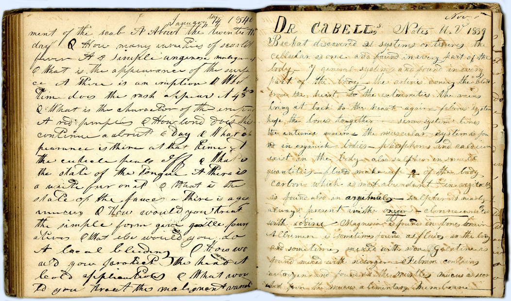
Student Notebook of Jethro Meriwether Hurt, 1839-1840. Hurt attended the University of Virginia for the 1839-40 session. See the top of the right hand page which refers to Dr. James L. Cabell, the Virginia anatomy professor at the time. Hurt graduated from the University of Pennsylvania Medical School in 1841. [2] University of Virginia Student Notebook Collection, Box 16, Folder 1, Historical Collections, Claude Moore Health Sciences Library, Charlottesville, Va.
The sporadic references to the Anatomical Theatre in minutes from the Board of Visitors meetings give some insight into the interior of the building and its use. A resolution made in July 1830, stipulated that the lecture room, which according to an early complaint lodged by Dr. Robley Dunglison was on the upper floor, [4] and dissection room be switched with the necessary removal and replacement of seats and the addition of a furnace and boiler in the new dissection room if necessary. In addition, the museum in the small basement room was to move to the large basement room, leaving the smaller to be used as a working room for Dr. Thomas Johnson, [5] the demonstrator of anatomy. [6]
Minutes from the next year’s meeting make it clear that not all these modifications had occurred as it was resolved, “That the Authority given to the Executive Committee at the last annual meeting of the Visitors to exchange the lecture and dissecting rooms at the Anatomical Theatre and to make alterations in those apartments having reference to such exchange be and the same is hereby revoked.” [7] Nevertheless, these minutes, dated less than six years after the opening of the University of Virginia, indicate that there was a substantial, functional basement, not just a charnel on the lower level as indicated in Thomas Jefferson’s design.
Outside the Theatre, it was agreed in 1831 that the contiguous kitchen garden be removed so that a lawn and trees could be planted with the thought of a future botanical garden. [8] The plan for the botanical garden was evidently abandoned as two years later it was again resolved to remove the kitchen garden, but simply replace it with grass and trees. [9]
In 1831, it was also determined to address the dampness in the front, eastern side of the building that was affecting the museum, to add shutters to the windows of the museum room, and to prevent general entry to the Theatre by keeping the building locked. [10] Blocking visual access to the museum would come up several more times in the minutes of the Board of Visitors. Blinds were approved for the museum windows in 1833 and in 1843. [11] In 1833, it was again resolved to fix a leak in the eastern wall of the basement as well as put backs on the museum cases and add new cases if necessary. [12]
An entry in the journal of the Chairman of the Faculty in early April 1833 relates an unpleasant discovery in the University ice pond. The Chairman was informed that, “on drawing off the water from the ice pond, a day or two since, an anatomical subject had been found.” Upon communicating this news to the University Proctor, he was informed that a “skeleton had been sunk there” by students without the knowledge of the anatomy demonstrator, Dr. Johnson, for the purpose of “bleaching.” [13]
Just a little over a month later, another complaint was made to the Chairman by a student named J.B. Washington about the building he called the Anatomical Hall. He said that it had been “greatly neglected of late” by the servant who was to be attending it, and “it was now extremely offensive to the students occupying the dormitories in the vicinity.” The Chairman asked the Proctor to have the servant “remove all the offensive matter” and use “chloride of lime.” (While he was addressing this unpleasant subject, the Chairman also requested that the Proctor do everything in his power to keep all hogs off the lawn.) [14]
It is probably not a coincidence that in July of this same year the Board of Visitors agreed that a small brick building in a valley to the west of the Anatomical Theatre could be appropriated by the professor of anatomy and surgery as a “boiling house and receptacle for subjects immediately after dissection.” [15] It has been speculated that the building had been used as a workshop, storage shelter, or dormitory prior to 1833. [16]

Stiff Hall, 1910. This photo shows the original south wing with the cupola as well as an attached north wing built in the 1880s. [20] After the new School of Medicine opened in 1929, this building was used for the Extension Division, a precursor to the School of Continuing and Professional Studies. [21] It was torn down for the ground-breaking of Newcomb Hall in 1956. [22] Prints20654, Special Collections, University of Virginia Library, Charlottesville, Va.
That same year a Board of Visitors resolution was made to move the Museum which had been in the Anatomical Theatre basement up one floor into the dissecting room to free up space for another lecture room in the basement that was to be offered to other schools of the University. [23] Whether or not other schools were ever offered this room, or would have accepted given the other uses of the Theatre, is not clear. If one did, it could not have been for more than several sessions because three years later, in 1840, the minutes show a resolution to add a large stove to the middle of the basement lecture room to supplement smaller stoves along the sides to take away the winter chill, specifically for the professor of medicine and his students. [24]
Additional Anatomical Theatre Modifications
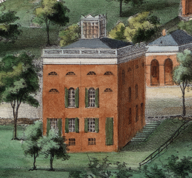
Detail of the Anatomical Theatre from View of the University … by E. Sachse & Co., 1856. A cupola was not part of Jefferson’s design. It was probably added after the 1837 resolution to raise the roof. This image clearly shows the cupola was present in 1856. Special Collections, University of Virginia Library, Charlottesville, Va.
In 1837, a resolution was made to raise the roof of the Theatre and cover it with slates. [25] The original roof with its skylight likely leaked as did so many of the early University buildings. It is possible the first roof was removed, but it is more likely that a second roof with a pitch was added over the first. Either way, Edward Sachse’s View of the University, Charlottesville & Monticello, taken from Lewis Mountain clearly shows that the cupola, absent in Jefferson’s original design, was present in 1856. It seems reasonable that the cupola was added when the new roof was installed. With its many panes of glass it would have functioned as a lantern during the day by sending shafts of light into the top floor of the Theatre.
Also in 1837, Dr. Augustus Warner, who had earlier waged a spirited battle for the use of the University wagon and horse to carry cadavers for dissection, suggested various improvements needed at the Anatomical Theatre. As a result, it was resolved to remove the brick pavement and vault that were constructed in front of the Theatre to span the space between the building and the road. They were to be replaced with a platform of plank to permit “a free passage of air under neath the platform between the parallel wall supporting the platform in front and the wall of the building.” In a process “recently discovered,” the wood was to have been “kiannized” [kyanized] or soaked in mercuric chloride. [26] This must have been proposed as a way to address the dampness problem referred to in prior years.
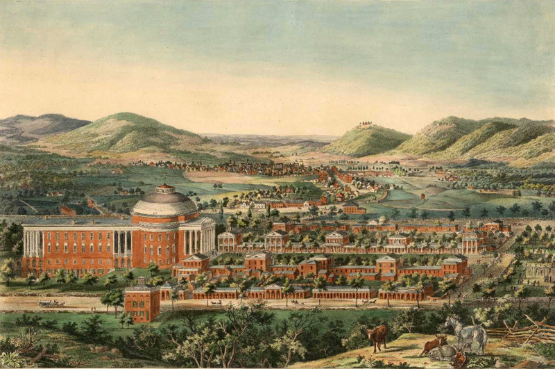
View of the University, Charlottesville & Monticello, taken from Lewis Mountain by E. Sachse & Co., published by Casimir Bohn, 1856. This shows the relation of the Anatomical Theatre in the left foreground to the rest of the University. The annex on the back of the Rotunda was added in 1853. Special Collections, University of Virginia Library, Charlottesville, Va.
Lighting was understandably an issue for the Anatomical Theatre. In 1843, the Proctor, under the direction of the anatomy professor, Dr. James L. Cabell, was authorized to make an alteration in the lecture room, “by removal of the present partition in the room, so as to admit light from the western window of the story, and by the formation of a new but smaller apartment for the reception of subjects for dissection in the South western angle of the building,” the corner that contained the charnel. [27] This mention of the reception of subjects indicates that some dissection continued to occur in the Anatomical Theatre after the building of the Dissecting Hall in 1837.
Interior modifications continued to be mentioned in the Board of Visitors minutes. Particularly, in 1850 it was recommended that the cupboards in the Museum should be altered to provide without delay an enlarged space for separate lecture rooms for Drs. Cabell and Davis because of increased student “numbers too large to be accommodated at lecture with such a regard to health or comfort as prudence demands.” [28]
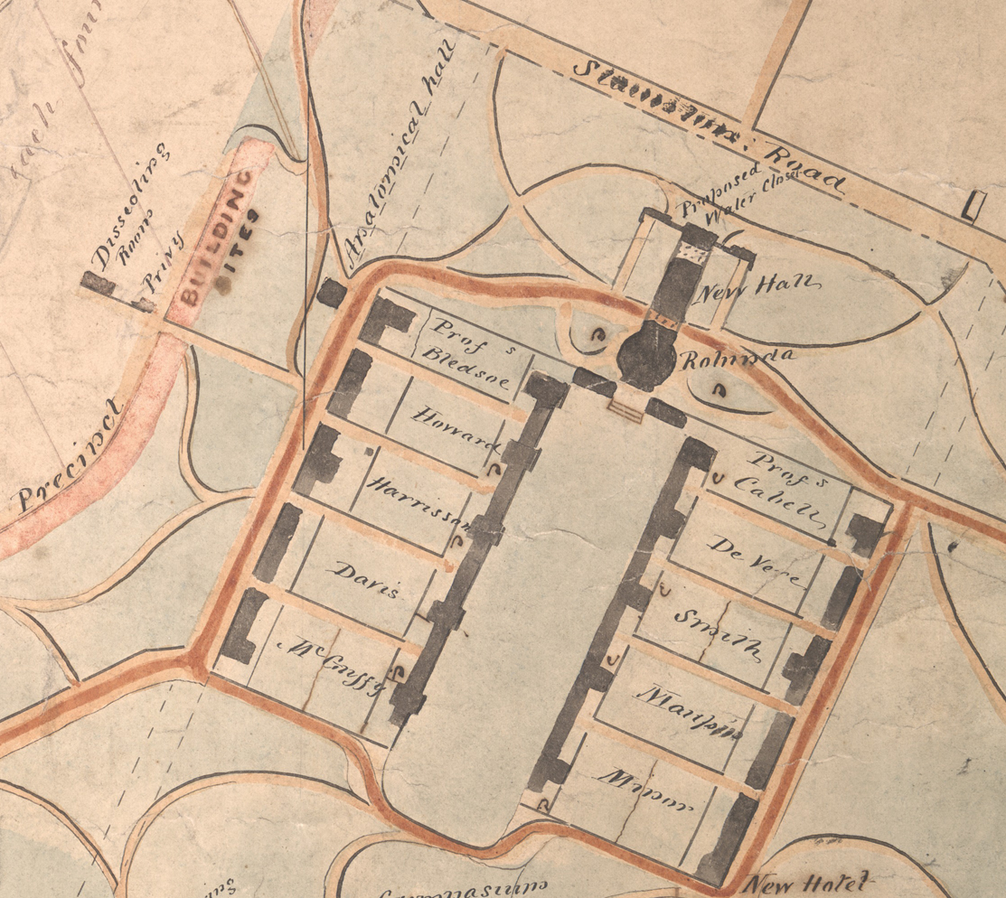
Detail of the Plan of University Cleared Land by
William Pratt 1858. University of Virginia Maps and Plats, 1856-1869, n.d., Accession # RG-31/1/2:2.532, Special Collections, University of Virginia, Charlottesville, Va.
The map to the left shows the University as it was in 1858. Clicking on it will show a larger version which makes it easier to see the relative placement of the Dissecting Rooms, also called Stiff Hall, and the Anatomical Hall or Theatre. Both are in the upper left quadrant. The pavilions are labeled with the names of the resident professors. Dr. Davis lived on the west side and Dr. Cabell on the east. The Rotunda is to the north of the pavilions and is connected to the New Hall, which was added in 1853. Later known as the Annex, it burned down in 1895.
Anatomical Theatre Museum and Plates
In 1837, Dr. Augustus Warner, who was instrumental in suggesting various improvements to the Anatomical Theatre itself, also made known to the Board of Visitors the condition of the anatomical plates then in use. [29] A Board resolution in 1837 to set aside $400 to purchase additional plates and another $400 to buy necessary anatomical preparations was evidently accomplished as the Catalogue for the 1842-1843 session of classes states the, “Anatomical and Pathological Museum … has been lately enriched with valuable and rare specimens selected in Paris by one of the Professors.” [30] Additionally in 1837, probably at the behest of Dr. Warner, the Board of Visitors minutes stipulated guidelines for the use of money that might be left from the $5 dissection fee collected from each student and the $100 given by the University for procuring subjects. Up to $50 could be spent for medical periodicals with any additional surplus to be used to purchase works on medical science. These periodicals and books were to be added to the collection of anatomical plates and books already housed in the Theatre. [31]
Board of Visitors minutes from the late 1840s indicate all was well, specifically in the anatomical museum: “every thing there bore the appearance of being well arranged & in good order; & the various preparations in the Museum in good condition & State of preservation.” [32] In 1850 Professor James L. Cabell, professor of zoology and comparative anatomy, was reimbursed for his purchase of plates and drawings related to those subjects. [33]
In 1856, the University employed Henry Scharf, an artist, illustrator, and actor, to make colored anatomical drawings for physiology and anatomy classes. He came to the University of Virginia after being recommended by the anatomy professor, Dr. A.E. Peticolas, at the Medical College of Virginia in Richmond. Dr. Peticolas suggested that the paintings be done, “as they do the scenes at theaters, with opaque colors on prepared cotton cloth … primed with whiting and glue“ and stretched on a frame. [34] In 1857 Scharf was paid $500 for his year’s work. At least $3,000 was paid over the course of six years, and the medical department consequently “accumulated an unequalled collection of plates, executed with an exquisite truth to nature, making them invaluable.” [35]
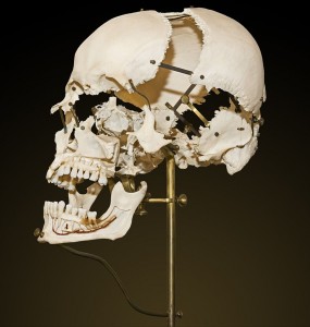
Beauchêne Skull. The Beauchêne skull is also called an exploded skull. Wikimedia Commons.
In the summer of 1859, Dr. Davis made an inventory and determined that the department of anatomy had 111 pictures in 9 waterproof cases; 2 articulated skeletons; 2 separated skulls; 3 remounted (a la Beauchêne) skulls, meaning ones that had been cleaned, disarticulated, and then reassembled with jointed supports so that bones could be studied both individually and in context; papier-mâché models of the brain, ear, eye, larynx, and throat; a series of fetal skeletons; additional separate bones; and numerous specimens, both natural and diseased, of organs of the body and dried, varnished or preserved in antiseptic liquid. His inventory of the department of materia medica included a cabinet with 250 specimens of medicines and a set of 98 engravings of medicinal plants. [36]
Informal Anatomical Drawings
The medical school purchased anatomical plates and drawings over the years, but either the Dissecting Hall or the Anatomical Theatre was home to a work of art by an unknown illustrator. Writing about his experience in the School of Medicine in 1876-1877 when he was a student, Dr. Paul Barringer described a mural that faded over time.
“In those days a great mural adorned the walls of the dissecting room. Nearly always there were one or more ‘academs’ in anatomy, artists or others after the artistic approach to the human figure, and one of these had drawn in permanent crayon a most surprising scene. The dissecting hall had been built fifty years earlier, and at the back end was a bricklined pit, some thirty or forty feet deep, into which was thrown all anatomical waste—particularly the bodies carved up in ‘surgery on the cadaver,’ along with many other fragments, for only the most stalwart bodies were boiled to make proper material for the work in osteology.
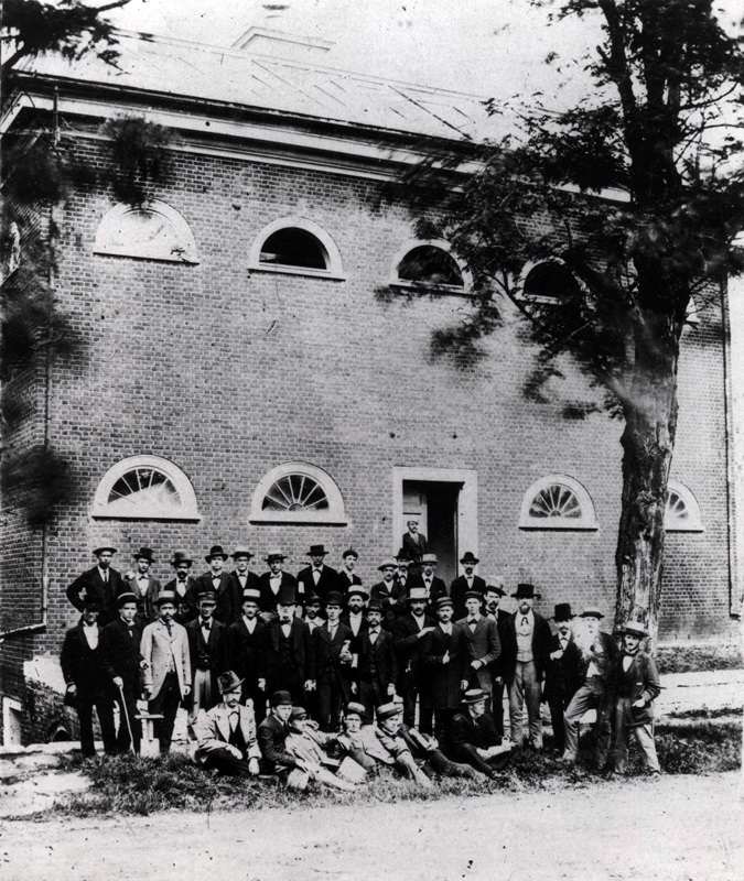
Anatomical Theatre with the Class of 1873. The class is standing in front of the Anatomical Theatre. The cupola, probably added after the 1837 resolution to raise the roof, is barely visible at the top of the building. Prints07390, Special Collections, University of Virginia Library, Charlottesville, Va.
The artist had taken as his subject the mouth of this great, nearly filled pit on Judgment Day, and he was an artist almost beyond compare with a sense of humor that literally lapped over. Great clouds emerged from the pit, and rose, and spread out even over the ceiling. On the ceiling section was Old Gabriel with his horn. Down at the mouth of the pit was mere anatomical debris, but a few feet higher the pieces began to get together with a full complement of human motive. Two thoraces were fighting over a pelvic bone. A little farther along a pair were matching femurs, to see which would suit best, but the real chef-d’oeuvre was in a cloud that ran under and rose up between two windows. Here were two lovers, the lady minus one tibia, seated on a rock while her inamorato ran back and forth from the melee, a tibia in each hand and half a dozen extra ones sticking out from the openings in his ribs where he had placed them for easy carriage. Believe it or not, he undoubtedly had a smile on his prognathic facial bones, and she seemed smugly demure as she sat with her sound leg crossed over a legless femur, patiently holding her detached foot in her hands.” This drawing was sacred, and although carefully preserved, time, tobacco smoke, and fumes from the boiling caldrons in the cellar were rapidly making away with it.” [37]
- University of Virginia, Board of Visitors. Minutes, June 28, 1850, 171, Retrieved from http://guides.lib.virginia.edu/bovminutes.
- Penn Medical Students: compiled from the January 1841 University Catalog. Retrieved from http://www.archives.upenn.edu/people/students/med/catalogs/catmedmat1841.html.
- University of Virginia. Catalogue of the Officers and Students of the University of Virginia, 1825-26 through 1846-47.
- Robley Dunglison to Arthur S. Brockenbrough, September 8, 1826. Papers of the Proctor of the University of Virginia, Box 6: Folder 655. RG-5/3/1.111, Special Collections, University of Virginia Library, Charlottesville, Va.
- The Demonstrator of Anatomy in 1927 was Dr. Thomas Johnson. He advanced to become a full professor in anatomy in 1832 and resigned in 1834. Blanton, Wyndham Bolling. Medicine in Virginia in the Nineteenth Century. Richmond: Garrett & Massie, 1933, 22.
- University of Virginia, Board of Visitors. Minutes, July 30, 1830, 28-29.
- University of Virginia, Board of Visitors. Minutes, July 11, 1831, 63.
- University of Virginia, Board of Visitors. Minutes, July 11, 1831, 63-64.
- University of Virginia, Board of Visitors. Minutes, July 10, 1833, 92.
- University of Virginia, Board of Visitors. Minutes, July 11, 1831, 63, 66.
- University of Virginia, Board of Visitors. Minutes, July 10, 1833, 92 and July 4, 1843, 73.
- University of Virginia, Board of Visitors. Minutes, July 10, 1833, 92.
- Journals of the Chairman of the Faculty 1827-1864, April 5, 1833, Box 1: Volume 4. RG-19/1/2.041, Special Collections, University of Virginia Library, Charlottesville, Va.
- Journals of the Chairman of the Faculty 1827-1864, May 14 1833, Box 1: Volume 4.
- University of Virginia, Board of Visitors. Minutes, July 10, 1833, 92.
- Fitz-Hugh, G. Slaughter. Anatomical Laboratory – Dissecting Hall 1833-1928, [manuscript], 1981, 3. Historical Collections & Services, Claude Moore Health Sciences Library, University of Virginia.
- University of Virginia, Board of Visitors. Minutes, August 17, 1837, 5.
- Fitz-Hugh, 1.
- Fitz-Hugh, 3-4.
- Barringer, Paul B., James M. Garnett and Rosewell Page, (Eds.). University of Virginia, Its History, Influence, Equipment and Characteristics. New York: Lewis Publishing Co., 1904, 109-110.
- Fitz-Hugh, 2; “100 Years of Partnership.” Columns: News from the School of Continuing & Professional Studies (Spring 2015) Retrieved from http://www.scps.virginia.edu/columns/view/100-years-of-partnership-Spring-2015.
- Fitz-Hugh, 4.
- University of Virginia, Board of Visitors. Minutes, August 17, 1837, 5.
- University of Virginia, Board of Visitors. Minutes, July 4, 1840, 47.
- University of Virginia, Board of Visitors. Minutes, August 17, 1837, 4.
- University of Virginia, Board of Visitors. Minutes, August 17, 1837, 4.
- University of Virginia, Board of Visitors. Minutes, July 4, 1843, 73.
- University of Virginia, Board of Visitors. Minutes, June 28, 1850, 171.
- University of Virginia, Board of Visitors. Minutes, August 17, 1837, 4.
- University of Virginia, Board of Visitors. Minutes, August 17, 1837, 4-5; University of Virginia. Catalogue of the Officers and Students of the University of Virginia, Session of 1842-1843. Richmond: Shepherd and Colin, 1843, 22.
- University of Virginia, Board of Visitors. Minutes, August 17, 1837, 6.
- University of Virginia, Board of Visitors. Minutes, June 25, 1848, 138.
- University of Virginia, Board of Visitors. Minutes, June 28, 1850, 171.
- A.E. Peticolas to J.S. Davis, January 30, 1856. Papers of John Staige Davis, 1839-1885, Box 1: Folder “Correspondence of John Staige Davis, Sr., 1851-1858.” MSS 3247, Special Collections, University of Virginia Library, Charlottesville, Va.
- Barringer, Paul B., James M. Garnett and Rosewell Page, (Eds.), 160.
- Inventory of the Department of Anatomy, the Department of Materia Medica, and the Department of Botany & Vegetable Physiology, June 23, 1859. Papers of John Staige Davis, 1840-1888, Box 3: Letterpress book. MSS 1912, 2842, Special Collections, University of Virginia Library, Charlottesville, Va.
- Barringer, Paul B. The Natural Bent. Chapel Hill: The University of North Carolina Press, 1949, 221-222.
Previous: “Subjects” for Anatomy Class
Next: The Post-Civil War Anatomical Theatre